Animal Study Finds Effects on Behavior, Brain Chemistry of Prenatal MDMA Exposure
Download PDF Version What is PDF?
Betsy Earp
Betsy Earp is a Contributing Writer for NIDA NOTES.
Source: NIDA NOTES, Vol. 19, No. 2, July, 2004
Public Domain
Table of Contents (TOC)
Article: Animal Study Finds Effects on Behavior, Brain Chemistry of Prenatal MDMA ExposureReferences
 Because women of childbearing age are among the population most involved
with MDMA (methylenedioxymethamphetamine, ecstasy), the consequences
of prenatal exposure to the drug are an important concern. Studies have
demonstrated subtle long-term behavioral abnormalities in animals exposed
to MDMA during the mid- to late gestational period. Now, NIDA-funded
researchers have documented marked behavioral abnormalities in rats
exposed to MDMA during early in utero life. Moreover, the researchers
correlated the exposed rats’ abnormal behavior at 21 days of age with
alterations in brain neurotransmitter systems that were passing through
a critical early developmental stage at the time of their in utero exposure.
These findings strengthen the evidence, which has been conflicting to
date, that prenatal MDMA exposure has an impact on neurochemical development.
"This basic study is a significant step toward understanding the consequences for the offspring of MDMA exposure in early pregnancy," says Dr. Pushpa Thadani of NIDA’s Division of Neuroscience and Behavioral Research. "More studies are needed, as well as monitoring of children prenatally exposed to MDMA."
Dr. Jack Lipton at Rush University Medical Center in Chicago injected pregnant albino rats with MDMA, 15 mg/kg of body weight, equivalent to a human recreational dose, twice daily when the animals were 14 to 20 days pregnant. A control group of pregnant albino rats received 1 mL/kg saline during the same pregnancy interval. During this time gestating rats pass through a critical brain developmental stage, corresponding to a stage that occurs during early pregnancy in humans, in which the dopamine and serotonin brain neurotransmitter systems first take shape. Dopamine is involved in motivated behaviors, including attention, sex, and drug taking, while serotonin helps regulate mood, sleep, and appetite.
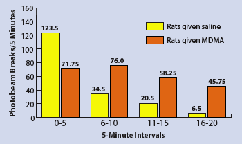
After the rat pups were born, the researchers tested them at several ages for signs of structural changes in the dopamine and serotonin neurotransmitter systems and at postnatal day 21 for behavioral deficits. In the behavioral tests, they examined the rats’ adaptation to a new environment by introducing the animals into an unfamiliar cage that was equipped to monitor the animals’ ambulatory and fine motor movements. They found that at 21 days of age, the rats prenatally exposed to MDMA adapted more slowly to the novel environment and exhibited significantly more activity than the unexposed rats. The exposed rats maintained a 35-percent increase over their normal activity level throughout the 20-minute trial. In contrast, the activity level of rats in the control group fell off precipitously after the first 5 minutes of the trial as they adjusted to the new cage.
Rats Prenatally Exposed to MDMA Are More Active Than Unexposed Rats
 When 21 days old, rats prenatally exposed to MDMA and those from a control group were each placed in a new cage, equipped with photobeams to monitor their activity. MDMA-exposed rats were seven times as active as the control group rats by trial’s end.
 To investigate for possible MDMA-related abnormalities in brain chemistry and structure, the researchers measured the levels of dopamine and serotonin and their metabolites in selected brain regions in MDMA-exposed and unexposed male and female rat offspring at different postnatal ages. Although nothing was remarkable about these measurements at the 3-day mark, distinctions between the animals emerged at 21 days. At that age, rats that had been prenatally exposed to MDMA showed significant reductions in dopamine and serotonin metabolites and turnover in the nucleus accumbens and striatum. The researchers hypothesize that these changes reflect either a decrease in dopamine or serotonin release, a reduction in enzymatic activity, or an increase in dopamine neuron innervation.
The researchers also sought to determine whether prenatal exposure to MDMA had altered the dopamine nerve fiber density in several regions of the brain. They found that fiber counts in the frontal cortex of the prenatally exposed 21-day-old rats had increased 502 percent more than counts seen in controls—of concern, as abnormal or excessive connections in the frontal cortex may result in aberrant signaling, possibly leading to abnormal behavior.
In summary, reductions in prenatally exposed rats’ dopamine and serotonin metabolism, an inability to adjust to a new environment, and increases in dopamine fiber density demonstrate for the first time that prenatal MDMA exposure in rats may result in both behavioral and correlating neurochemical alterations. Dr. Lipton observes, "The health risks that MDMA exposure poses to fetuses are not fully known, and continued research into the consequences of exposure and monitoring of prenatally exposed children is certainly warranted."
Koprich, J.B.; Chen, E.Y.; Kanaan, N.M.; Campbell, N.G.; Kordower, J.H.; and Lipton, J.W. Prenatal 3,4-methylenedioxymethamphetamine (ecstasy) alters exploratory behavior, reduces monoamine metabolism, and increases forebrain tyrosine hydroxylase fiber density of juvenile rats. Neurotoxicology and Teratology 25(5):509-517, 2003.


