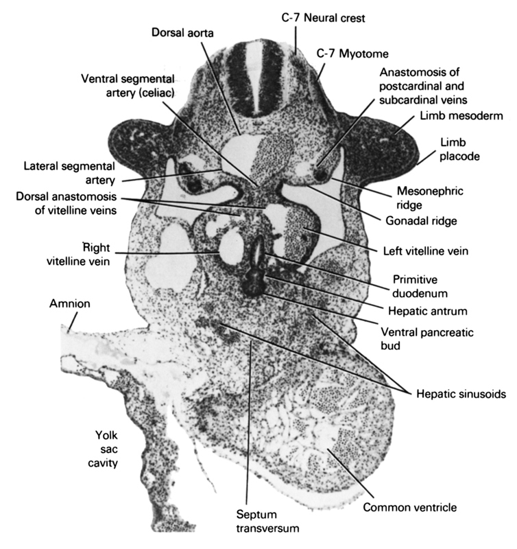
A section through the C-5 neural crest and the primary interventricular foramen.
Observe:
1. The interventricular sulcus separating the primitive right and left ventricles and the caudalmost extent of the sinus venosus in the septum transversum.
2. The hepatic trabeculae invading the septum transversum.
3. The anastomosis of the vitelline veins ventral to the primitive duodenum and the umbilical veins in the lateral body wall.
4. The dorsal pancreatic bud from which the body and tail of the pancreas develop.
5. The communication of the hepatocardiac veins with the venous channels between the hepatic trabeculae.
Keywords: C-7 myotome, C-7 neural crest, amnion, anastomosis between postcardinal and subcardinal veins, common ventricle, dorsal anastomosis of vitelline veins, dorsal aorta, gonadal ridge, hepatic antrum, hepatic sinusoids, lateral segmental artery, left vitelline (omphalomesenteric) vein, limb mesoderm, limb placode, mesonephric ridge, primitive duodenum, right vitelline (omphalomesenteric) vein, septum transversum, ventral pancreatic bud, ventral segmental artery (celiac), yolk sac cavity
Source: Atlas of Human Embryos.
