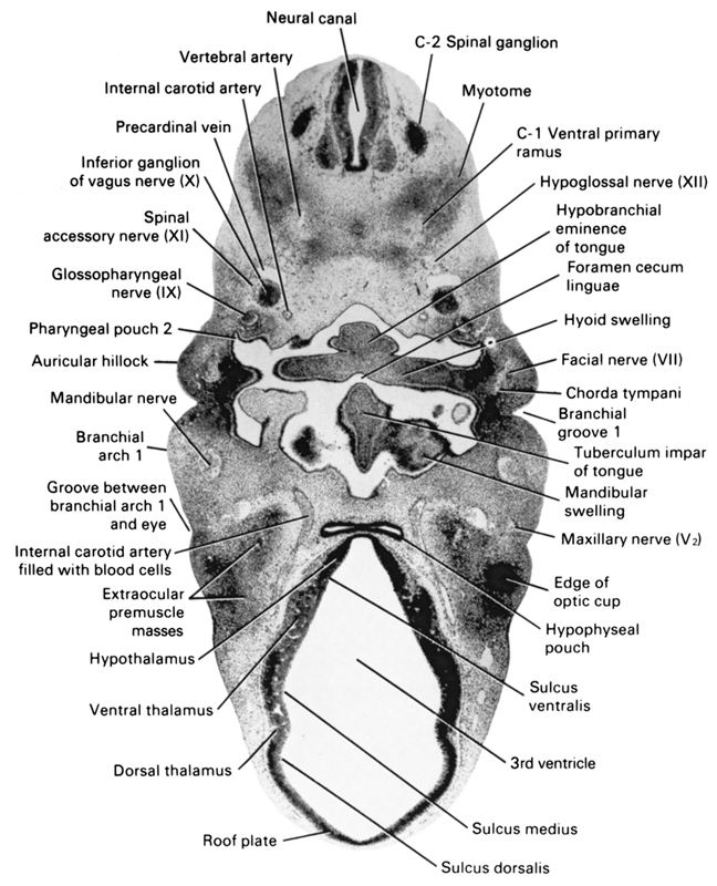
A section through the C-2 spinal ganglion and dorsal surface of the tongue.
Observe:
1. The inferior ganglion of the vagus medial to the spinal accessory nerve.
2. The maxillary and mandibular nerves in the first branchial arch.
3. The facial and glossopharyngeal nerves on each side of the second pharyngeal pouch.
4. The chorda tympani nerve arising from the facial nerve.
5. The internal carotid artery flanking each side of the hypophyseal pouch.
Keywords: C-1 ventral primary ramus, C-2 spinal ganglion, auricular hillock, branchial arch 1, branchial groove 1, chorda tympani, dorsal thalamus, edge of optic cup, extra-ocular premuscle masses, facial nerve (CN VII), foramen cecum linguae, glossopharyngeal nerve (CN IX), groove between branchial arch 1 and eye, hyoid swelling, hypobranchial eminence of tongue, hypoglossal nerve (CN XII), hypophyseal pouch, hypothalamus, inferior ganglion of vagus nerve (CN X), internal carotid artery, internal carotid artery filled with blood cells, mandibular nerve, mandibular swelling, maxillary nerve (CN V₂), myotome, neural canal, pharyngeal pouch 2, precardinal vein, roof plate, spinal accessory nerve (CN XI), sulcus dorsalis, sulcus medius, sulcus ventralis, third ventricle, tuberculum impar of tongue, ventral thalamus, vertebral artery
Source: Atlas of Human Embryos.
