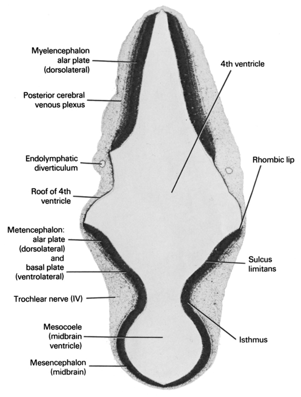
A section through the mesencephalic isthmus and tip of the endolymphatic diverticulum.
Observe:
1. The alar plate of the myelencephalon with the posterior cerebral venous plexus on its surface.
2. The alar and basal plates of the metencephalon separated by the sulcus limitans.
3. The rhombic lip of the alar plate where the cerebellum will later develop.
4. The minute trochlear nerve coursing ventrally on each side of the isthmus.
Keywords: endolymphatic diverticulum, isthmus, mesencephalon (midbrain), mesocoele (midbrain ventricle), metencephalon: alar plate (dorsolateral) and basal plate (ventrolateral), myelencephalon alar plate (dorsolateral), posterior cerebral venous plexus, rhombencoel (fourth ventricle), rhombic lip, roof of rhombencoel (fourth ventricle), sulcus limitans, trochlear nerve (CN IV)
Source: Atlas of Human Embryos.
