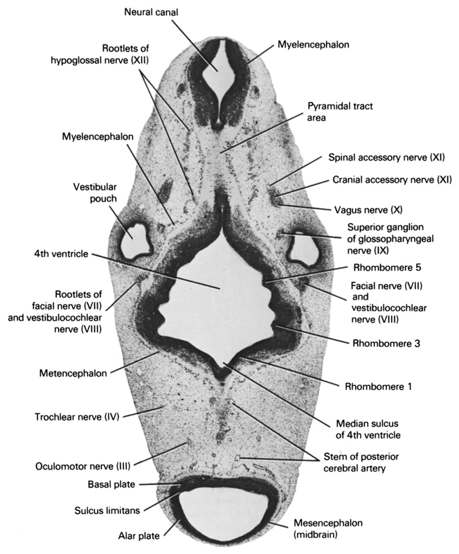
A section through the rootlets of the facial, vestibulocochlear and hypoglossal nerves.
Observe:
1. The pyramidal tract area on the ventral side of the myelencephalon.
2. The rhombomeres in the wall of the fourth ventricle.
3. The union of the cranial accessory nerve with the vagus nerve.
4. The superior ganglion of the glossopharyngeal nerve.
Keywords: alar plate(s), basal plate, cranial accessory nerve (CN XI), facial nerve (CN VII) and vestibulocochlear nerve (CN VIII) , median sulcus of 4th ventricle, mesencephalon (midbrain), metencephalon, myelencephalon, neural canal, oculomotor nerve (CN III), pyramidal tract area, rhombencoel (fourth ventricle), rhombomere 1, rhombomere 3, rhombomere 5, root of facial nerve (CN VII), root of hypoglossal nerve (CN XII), root of vestibulocochlear nerve (CN VIII), spinal accessory nerve (CN XI), stem of posterior cerebral artery, sulcus limitans, superior ganglion of glossopharyngeal nerve (CN IX), trochlear nerve (CN IV), vagus nerve (CN X), vestibular pouch
Source: Atlas of Human Embryos.
