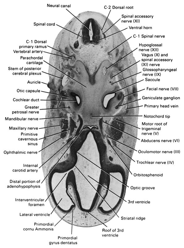
A section through the base of the developing skull (cranial tip of notochord and parachordal cartilage).
Observe:
1. The vessels and cranial nerves lateral to the distal portion of the adenohypophysis.
2. The three divisions of the trigeminal nerve; the ophthalmic, maxillary and mandibular nerves.
3. The geniculate ganglion on cranial nerve VII and its position lateral to the saccule and cochlear duct.
4. The junction of the primary head vein with the stem of the posterior cerebral plexus.
5. The relation of the C-1 spinal nerve to the vertebral artery.
Keywords: C-1 dorsal primary ramus, C-1 spinal nerve, C-2 dorsal root, abducens nerve (CN VI), auricle, cochlear duct, distal portion of adenohypophysis, facial nerve (CN VII), geniculate ganglion (CN VII), glossopharyngeal nerve (CN IX), greater petrosal nerve, hypoglossal nerve (CN XII), internal carotid artery, interventricular foramen, lateral ventricle, mandibular nerve (CN V₃), maxillary nerve (CN V₂), motor root of trigeminal nerve (CN V), neural canal, notochord tip, oculomotor nerve (CN III), ophthalmic nerve, optic groove, orbitosphenoid, otic capsule, parachordal cartilage, primary head vein, primitive cavernous sinus, primordial cornu Ammonis, primordial gyrus dentatus, roof of 3rd ventricle, saccule(s), spinal accessory nerve (CN XI), spinal cord, stem of posterior cerebral plexus, striatal ridge, third ventricle, trochlear nerve (CN IV), vagus nerve (CN X), ventral horn, vertebral artery
Source: Atlas of Human Embryos.
