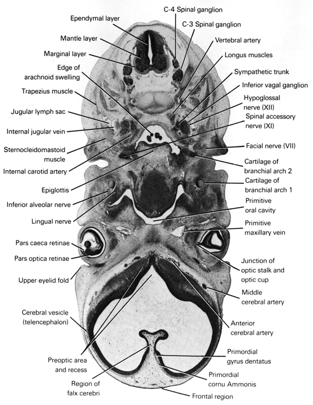
A section through the C-4 spinal ganglion and the preoptic area of the telencephalon.
Observe:
1. The junction of the optic stalk with the optic cup.
2. The two parts of the retina: pars caeca retinae and pars optica retinae.
3. The region of the falx cerebri between the cerebral vesicles.
4. The first and second branchial arch cartilages.
5. The three layers of the spinal cord in the midcervical region.
Keywords: C-3 spinal ganglion, C-4 spinal ganglion, anterior cerebral artery, cartilage of branchial arch 2, cerebral vesicle (telencephalon), edge of arachnoid swelling, ependymal layer, epiglottis, facial nerve (CN VII), falx cerebri region, frontal region, hypoglossal nerve (CN XII), inferior alveolar nerve, inferior ganglion of vagus nerve (CN X), internal carotid artery, internal jugular vein, jugular lymph sac, junction of optic stalk and optic cup, lingual nerve, longus muscles, mantle layer, marginal layer, middle cerebral artery, pars caeca retinae, pars optica retinae, pharyngeal arch 1 cartilage (Meckel), preoptic area and recess, primitive maxillary vein, primitive oral cavity, primordial cornu Ammonis, primordial gyrus dentatus, spinal accessory nerve (CN XI), sternocleidomastoid muscle, sympathetic trunk, trapezius muscle, upper eyelid fold, vertebral artery
Source: Atlas of Human Embryos.
