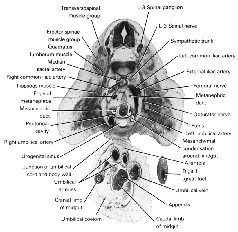
A section through the L-3 spinal ganglion and the caudal edge of the metanephros.
Observe:
1. The appendix represented as an outpouching of the caudal limb of the midgut.
2. The caudal edge of the junction of the umbilical cord and the ventral body wall.
3. The primordium of the first digit of the foot.
4. The obturator nerve coursing medial to the pubis of the developing pelvic bone.
5. The three terminal branches of the aorta: right and left common iliac and median sacral arteries.
Keywords: L-3 spinal ganglion, L-3 spinal nerve, allantois, appendix, caudal limb of midgut, cranial limb of midgut, digit 1 (great toe), edge of metanephros, erector spinae muscle group, external iliac artery, femoral nerve, iliopsoas muscle, junction of umbilical cord and body wall, left common iliac artery, left umbilical artery, median sacral artery, mesenchymal condensation around hindgut, mesonephric duct, metanephric duct, obturator nerve, peritoneal cavity, pubis, quadratus lumborum muscle, right common iliac artery, right umbilical artery, sympathetic trunk, transversospinal muscle group, umbilical arteries, umbilical coelom, umbilical vein, urogenital sinus
Source: Atlas of Human Embryos.
