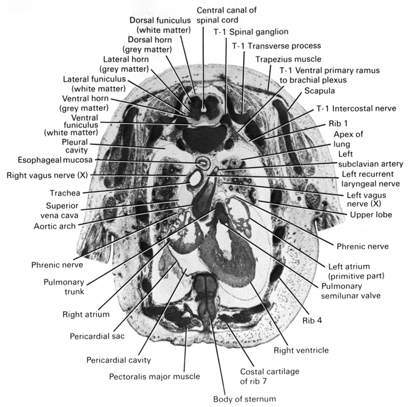
A section through the caudal part of the sternum, cranial part of the heart and lungs and T-1 spinal ganglion.
Observe:
1. The pulmonary semilunar valve separating the right ventricle and pulmonary trunk.
2. The aortic arch passing obliquely to the left of the trachea and esophagus.
3. The left vagus nerve coursing between the aortic arch and the left lung with its recurrent laryngeal branch looping around the arch and ascending to the larynx.
4. The origin of the left subclavian artery from the aortic arch.
5. The subdivisions of the grey and white matter in the spinal cord.
Keywords: T-1 intercostal nerve, T-1 spinal ganglion, T-1 ventral primary ramus to brachial plexus, apex of lung, arch of aorta, body of sternum, central canal of spinal cord, dorsal funiculus (white matter), dorsal horn (grey matter), esophageal mucosa, lateral funiculus (white matter), lateral horn (grey matter), left atrium (primitive part), left recurrent laryngeal nerve, left subclavian artery, left vagus nerve (CN X), pectoralis major muscle, pericardial cavity, pericardial sac, phrenic nerve, pleural cavity, pulmonary semilunar valve, pulmonary trunk, rib 1, rib 4, rib 7 (costal cartilage), right atrium, right vagus nerve (CN X), right ventricle, scapula, superior vena cava, trachea, transverse process of T-1 vertebra, trapezius muscle, upper lobe of left lung, ventral funiculus (white matter), ventral horn (grey matter)
Source: Atlas of Human Embryos.
