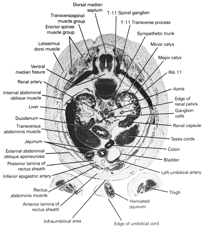
A section through the caudal edge of the umbilical cord, liver and duodenum and the T-11 spinal ganglion.
Observe:
1. The cut edge of the thigh on each side of the infraumbilical area of the abdominal wall.
2. The arrangement of the muscles and aponeuroses in the abdominal wall.
3. The origin of the right renal artery from the aorta.
4. The major and minor calyces of the renal pelvis.
5. The ganglion cells ventral to the aorta between the kidneys.
Keywords: T-11 spinal ganglion, T-11 transverse process, anterior lamina of rectus sheath, aorta, colon, dorsal median septum, duodenum, edge of renal pelvis, edge of umbilical cord, erector spinae muscle group, external abdominal oblique aponeurosis, ganglion cells, herniated jejunum, inferior epigastric artery, infra-umbilical area, internal abdominal oblique muscle, jejunum, latissimus dorsi muscle, left umbilical artery, liver, major calyx, minor calyx, posterior lamina of rectus sheath, rectus abdominis muscle, renal artery, renal capsule, rib 11, sympathetic trunk, testis, thigh, transversopinal muscle group, transversus abdominis muscle, urinary bladder, ventral median fissure
Source: Atlas of Human Embryos.
