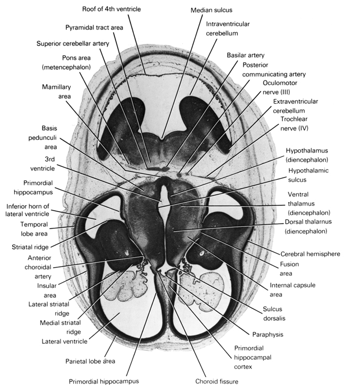
A section through the cerebral hemispheres (insular area and parietal and temporal lobe areas) and middle of the diencephalon and metencephalon.
Observe:
1. The striatal ridge in the floor of the lateral ventricle and the primordial hippocampus in the medial wall of the cerebral hemisphere.
2. The extension of the inferior horn of the lateral ventricle into the temporal lobe area.
3. The hypothalamic sulcus demarcating the thalamus from the hypothalamus.
4. The fusion area between the cerebral hemispheres and diencephalon.
5. The intra- and extraventricular parts of the cerebellum.
Keywords: anterior choroidal artery, basilar artery, basis pedunculi area, cerebral hemisphere, choroid fissure, diencoel (third ventricle), dorsal thalamus (diencephalon), extraventricular cerebellum, fusion area, hypothalamic sulcus, hypothalamus (diencephalon), inferior horn of lateral ventricle, insular area, internal capsule area, intraventricular cerebellum, lateral striatal ridge, lateral ventricle, mamillary area, medial striatal ridge, median sulcus, oculomotor nerve (CN III), paraphysis, parietal lobe area, pons region (metencephalon), posterior communicating artery, primordial hippocampal cortex, primordial hippocampus, pyramidal tract area, roof of rhombencoel (fourth ventricle), striatal ridge, sulcus dorsalis, superior cerebellar artery, temporal lobe area, trochlear nerve (CN IV), ventral thalamus (diencephalon)
Source: Atlas of Human Embryos.
