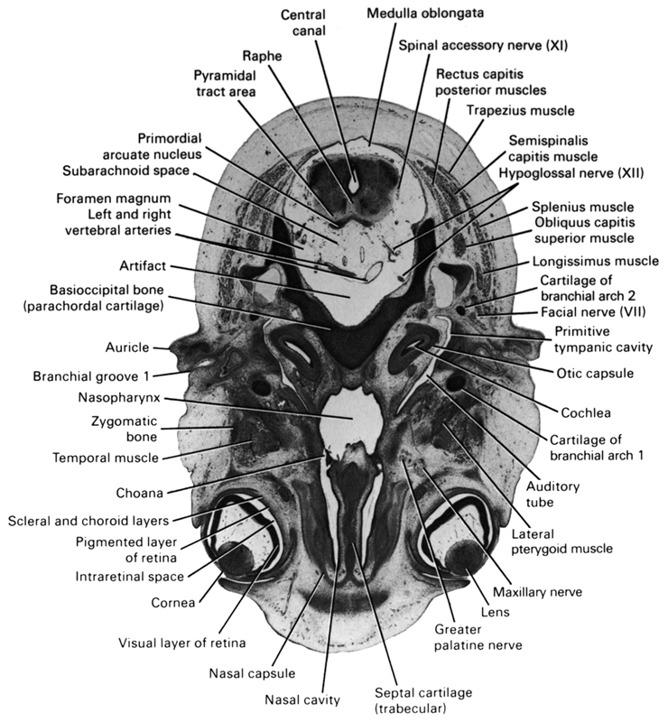
A section through the middle of the eye and caudal part of the medulla oblongata.
Observe:
1. The layers of the eye.
2. The muscles of mastication between the eye and the auditory tube (pouch 1).
3. The choana through which the nasal cavity communicates with the nasopharynx.
4. The relation of the first and second arch cartilages to the middle ear.
5. The hypoglossal nerve in the subarachnoid space.
Keywords: artifact(s), auditory tube, auricle, basi-occipital bone (parachordal cartilage), cartilage of pharyngeal arch 2, central canal, choana, cochlea, cornea, facial nerve (CN VII), foramen magnum, greater palatine nerve, hypoglossal nerve (CN XII), intraretinal space (optic vesicle cavity), lateral pterygoid muscle, left and right vertebral arteries, lens, longissimus muscle, maxillary nerve (CN V₂), medulla oblongata, nasal capsule, nasal cavity (nasal sac), nasopharynx, obliquus capitis superior muscle, optic part of retina, otic capsule, pharyngeal arch 1 cartilage (Meckel), pharyngeal groove 1, pigmented layer of retina, primitive tympanic cavity, primordial arcuate nucleus, pyramidal tract area, raphe, rectus capitis posterior muscle, scleral and choroid layers, semispinalis capitis muscle, septal cartilage (trabecular), spinal accessory nerve (CN XI), splenius muscle, subarachnoid space, temporalis muscle, trapezius muscle, zygomatic bone
Source: Atlas of Human Embryos.
