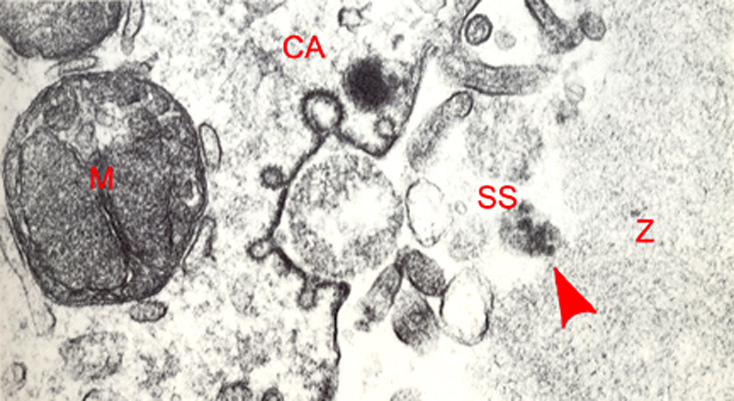
High power EM of a stage 1 embryo in vitro, 24 hours post-insemination showing delayed cortical granule release (original magnification x66,290). The simultaneous exocytosis of a cortical granule and formation of endocytotic caveolae (CA) are evident in this pronuclear embryo. The cell membrane at the site of the exocytosis is dense and striated. The nearby mitochondrion (M) is associated with smooth endoplasmic reticulum vesicles. The released products of cortical granules (arrow head) are visible in the subzonal space (SS).
Z = zona (capsula) pellucida
From: Sathananthan et al., 1986.
Keywords: cortical granule(s), endocytotic caveolae, exocytosis, mitochondria, smooth endoplasmic reticulum vesicles, subzonal space, zona pellucida
Source: The Virtual Human Embryo.
