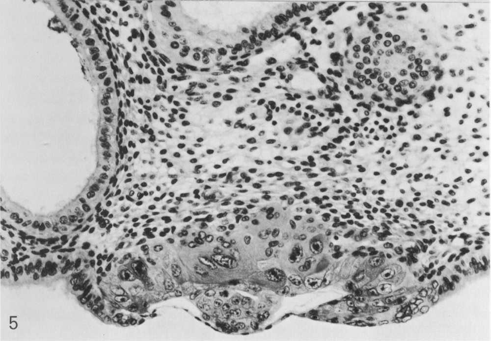
Section through the middle of the embryo. The amniotic cavity and bilaminar embryonic disc can be seen. The transition from the thin abembryonic trophoblast to the thick, solid layer at the embryonic pole is evident. Large multinucleated masses of syncytiotrophoblast project into the endometrial stroma. A dilated endometrial gland is cut through at the left-hand side of the photomicrograph.
Fig. 5. O'Rahilly and Müller, 1987.
Click on the picture to view the full-sized image.
Keywords: abembryonic trophoblast, amniotic cavity, bilaminar embryonic disc, endometrial gland, syncytiotrophoblast
Source: The Virtual Human Embryo.

