Dorsal Right Lateral Ventral Left Lateral |
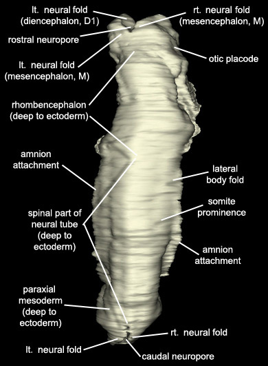
Reconstruction of embryo 6344's anatomy. Note that for this reconstruction of the anatomy the sections were realigned to straighten the twists and curves in the embryo's shape.
Keywords: amnion attachment, caudal eminence, caudal intestinal portal, caudal neuropore, cephalic intestinal portal, cephalic neuropore, cloacal membrane region, communication between peritoneal cavity and extra-embryonic coelom, connecting stalk, head fold region, heart prominence, lateral body fold, left neural fold, left neural fold [diencephalon (D1)], left neural fold [mesencephalon (M)], left otic placode, left umbilical vein, mandibular prominence of pharyngeal arch 1, maxillary prominence of pharyngeal arch 1, midgut, midgut endoderm, notochord, olfactory placode, optic sulcus, otic placode, paraxial mesoderm (deep to ectoderm), pharyngeal groove 1, rhombencephalon (deep to ectoderm), right neural fold, right neural fold [diencephalon (D1)], right neural fold [mesencephalon (M)], right umbilical vein, somite prominence, spinal part of neural tube (deep to ectoderm), stomodeum, tail fold region, umbilical vesicle wall attachmentSource: The Virtual Human Embryo.
