


0 μm
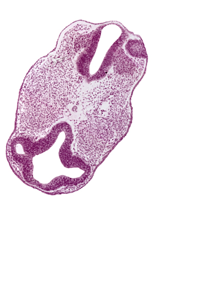
Carnegie Embryo #8943 | Location: 02-02-05
Keywords: cephalic edge of dorsal aorta, cephalic edge of notochord, cephalic edge of otic vesicle, communication between intraretinal space (optic vesicle cavity) and prosencoel (third ventricle), diencephalon (D1), diencephalon (D2), facio-vestibulocochlear neural crest (CN VII and CN VIII), intraretinal space (optic vesicle cavity), optic vesicle (D1), prosencoel (third ventricle), rhombencephalon (Rh. 3), rhombencephalon (Rh. 4), vascular plexus
Source: The Virtual Human Embryo.