Skip Navigation

Prenatal Form and Function – The Making of an Earth Suit

  
[Help]

Unit 7:   6 to 7 Weeks

  Closer Look: 
  Applying the Science: 
Play Movie - Human embryo 6 to 7 weeks
Movie 7.1 - 6 to 7 Weeks
Embryo nose, nasal plugs, 7 weeks 3 days
Figure 7.1 - Nasal Plugs Visible
The Biology of Prenatal Development DVD
Copyright © 2006 EHD, Inc. All rights reserved.


The embryo has brainwaves by 6 weeks, 2 days!

From 6 to 6½ weeks, the cerebral vesicles will double in size.1 Individualized brainwaves recorded via electroencephalogram (e-lek’tro-en-sef’a-lo-gram), or EEG, have been reported as early as 6 weeks, 2 days.2


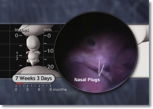
Also by 7 weeks, cell groupings resembling taste buds appear on the tongue3 and hiccups begin.4 Nasal plugs are prominent at this time and will persist for another 6 weeks or so.

Play Movie - Brainwaves
Movie 7.2 - Brainwaves
Play Movie - Hiccups
Movie 7.3 - Hiccups


By 7 weeks, the heart has 4 chambers.5

The pacemaker center is already well-established in the right atrium.6 The embryonic heart rate peaks at 7 weeks and now beats approximately 167-175 times per minute.7 This rate gradually declines to about 140 beats per minute at birth.8

Play Movie - The Maturing Heart
Movie 7.4 - The Maturing Heart
Adult Heart, 4 (four) chambers
Figure 7.2 - Adult Heart
Though the blood flow pattern before birth differs from the pattern after birth, the embryo has all 4 chambers similar to those seen in this adult heart.
LifeART
Copyright © 2002 Lippincott, Williams & Wilkins.


By 6½ weeks, the elbows are distinct and the embryo begins moving both hands. The fingers are also starting to separate as evidenced by the appearance of notching between the digital rays.9

The foot plate and ankle also emerge while toes and metatarsal (met’a-tar’sal) bones begin to form in the feet. Joint development is underway10 and the onset of primary muscle fiber formation indicates the embryo’s muscles are growing.11

Play Movie - Hand Movement
Movie 7.5 - Hand Movement
baby raising hand
Babies continue to fascinate themselves and us by moving their hands – an ability they have been practicing long before birth.


6 weeks 6 days Embryo foot plate, metatarsal bones
Figure 7.3 - Foot Plate
The Biology of Prenatal Development DVD
Copyright © 2006 EHD, Inc. All rights reserved.
Play Movie - Hand Plates
Movie 7.6 - Hand Plates
 

The hands and feet change dramatically between 6 and 7 weeks as separate fingers and toes begin to emerge. At 6 weeks the hand plates develop a subtle flattening between the digital rays. By 6 weeks, 2 days the hand takes on a polygon shape; prominent notches appear between the digital rays by 7 weeks and individual fingers are fully separated by 7½ weeks.12

Embryo Hand Development Progression: polygon shaped, notched fingers, fingers free
Figure 7.4 - Hand Development Progression (A, B, C)
Notice the hand’s progression from a polygon shape with digital rays, to definite notches between fingers, and finally to distinct fingers. The feet follow the same development pattern a few days later.
The Biology of Prenatal Development DVD
Copyright © 2006 EHD, Inc. All rights reserved.


Bone formation begins between 6 and 7 weeks, starting with the clavicle, or collar bone, and the upper and lower jaw. This process is called ossification.13


No Bones About It

Healthy food

Ossification is the process by which bones harden. But how does it happen? Basically, either existing membranes or cartilage (that firm connective tissue found in your ears and the tip of your nose) begin to harden through repeated deposits of calcium and phosphorus. Cells called osteoblasts form a bony framework. Osteoblasts then produce enough small chunks of bone to cover this framework, creating a solid bone.

Dairy products and many dark green veggies (such as spinach) are high in calcium which helps build bones providing the shape and structure of our body.

- Stedman's Medical Dictionary, Ed. Marjory Spraycar, et al., 26th ed. (Baltimore: Williams and Wilkins, 1995), 1266.
- Keith L. Moore and T. V. N. Persaud, The Developing Human, Clinically Oriented Embryology, 6th ed. (Philadelphia: W. B. Saunders, 1998), 408-411.

Play Movie - Bone Formation
Movie 7.7 - Bone Formation
Old Woman
Did you know the oldest bones in your body are your collar bone and jaw? These bones begin to develop in the womb by 7 weeks after fertilization.


Also, by 7 weeks, the embryo moves the legs and exhibits a startle response.14

Play Movie - Startle Response
Movie 7.8 - Startle Response
Skateboarding
Joints, muscles, and bones work together to enable the body to achieve feats like this.


Open PDF version of FIG 7.5, External Features and Nervous System of 18mm Embryo
Figure 7.5 - External Features (A) and Nervous System (B) of 18mm Embryo [PDF version of FIG 7.5]
From Gasser RF, 1975, 165. Atlas of Human Embryos. Copyright © 1975 RF Gasser, PhD. All rights reserved.


smiling nurse
A subset of proteins called antibodies fight infection and help prevent us from getting sick. However, sometimes our bodies need some outside help.
 

The immune system is maturing, as evidenced by the presence of B-lymphocytes in the liver.15 After birth and relocation away from the protection of the womb, these lymphocytes will produce proteins called antibodies to fight infection.

By 7 weeks, the ovaries appear in the female embryo.16 In the male embryo, a gene on the Y-chromosome produces a substance causing the testes to begin to differentiate.17

back to top
Take a written quiz about this unit. Take a visual quiz about this unit.
6 to 7
Weeks
Footnotes

1 Marin-Padilla, 1983. 25.
2 Bernstine, 1961. 63, 66; Bernstine et al., 1955. 623-30; Borkowski and Bernstine, 1955. 363. (cited by Bernstine, 1961, 63 & 66; O'Rahilly and Müller, 1999a, 195; and van Dongen and Goudie, 1980, 193.); Hamlin, 1964. 113; O'Rahilly and Müller, 1999a. 195; van Dongen and Goudie, 1980. 193.
3 Bradley and Stern, 1967. 750-751; Lawless, 1985. 579; Miller, 1982. 169; Mistretta and Bradley, 1975. 80; Ross and Nijland, 1997. 356.
4 de Vries et al., 1982. 305, 311; Visser et al., 1992. Figure 1, 176.
5 Cooper and O'Rahilly, 1971. 292; James, 1970. 214; Jordaan, 1979. 214; Moore, 1977. 298; Streeter, 1948. 192; Vernall, 1962. 23.
6 James, 1970. 214.
7 van Heeswijk et al., 1990. 153.
8 Leeuwen et al., 1999. 265.
9 O'Rahilly and Müller, 2001. 221; Streeter, 1948. 187.
10 Moore and Persaud, 2003. 385.
11 Edom-Vovard et al., 1999. 191.
12 Moore et al., 1994. Table 2-1, 61.
13 Carlson, 2004. 189; O'Rahilly and Gardner, 1972. 293; O'Rahilly and Gardner, 1975. 19; O'Rahilly and Müller, 2001. 385; Sperber, 1989. 122, 147; Spraycar, 1995. 1266.
14 de Vries et al., 1988. 96; Goodlin, 1979. D-128; Visser et al., 1992. Figure 1, 176.
15 Cunningham FG et al., 2001. 148.
16 O'Rahilly and Müller, 2001. 320.
17 Moore et al., 1994. 213, 216; Waters and Trainer, 1996. 15-16.