Developmental Stages in Human Embryos
Go to Stage: Intro 1 2 3 4 5 6 7 8 9 10 11 12 13 14 15 16 17 18 19 20 21 22 23
Stage 21
Page 256
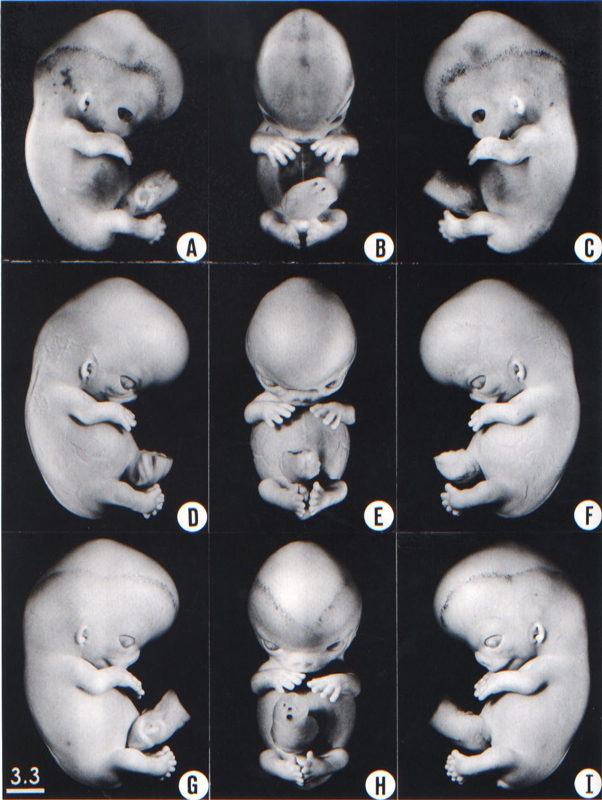
Fig. 21-1. Photographs of three embryos belonging to stage 21. The superficial scalp vascular plexus of the head is plainly visible in several of these views, such as A. It has spread to a level more than halfway between an eye-ear line and the vertex of the head. The fingers are longer and show an early phase in the development of touch pads. These Tastballen are shown at higher magnification by Cummins (1929) at stage 20 (his fig. 7) and stage 22 (his fig. 8.) The hands are flexed at the wrists and are approaching each other over the cardiac region. The lower limbs are curving toward the median plane, and toes of the two feet make contact with each other in some specimens. Top row, No. 4090. Middle row, No. 8553. Bottom row, No 7392. All views are at the same magnification.
Page 257SIZE AND AGE
Most embryos of this stage measure 22–24 mm.
The age is believed to be approximately 52 postovulatory days.
EXTERNAL FORM
The superficial vascular plexus of the head has spread upward to form a line at somewhat more than half the distance from eye-ear level to the vertex.
The fingers are longer and extend further beyond the ventral body wall than they did in the previous stage. The distal phalangeal portions appear slightly swollen and show the beginning of tactile pads. The hands are slightly flexed at the wrists and nearly come together over the cardiac eminence. The feet are also approaching each other, and the toes of the two sides sometimes touch.
FEATURES FOR POINT SCORES
1. Cornea. Cells are beginning to invade the postepithelial layer, converting it into the substantia propria (Streeter, 1951, fig. 16).
2. Optic nerve. Remnants of ependyma are present and may extend along practically the whole length of the optic stalk. A hyaloid groove is visible at the bulbar end. A few nerve fibers are arriving at the brain.
3 Cochlear duct. The tip of the duct now points definitely "downward" (fig. 19-6).
4. Adenohypophysis. The thread-like stalk is beginning to be absorbed (fig. 19-7).
5. Vomeronasal organ. The opening of the sac is reduced in size, a short, narrow neck is present, and the end of the sac is expanded (fig. 19-9).
6. Submandibular gland. The duct has begun to form knob-like branches (fig. 19-10).
7. Metanephros. Spoon-shaped glomerular capsules are developing, but no large glomeruli are present yet (fig. 19-11).
8. Humerus. Cartilaginous phases 1-4 are present (Streeter, 1949, figs. 3, 19, and 20).
ADDITIONAL FEATURES
Heart. Some photomicrographs were reproduced by Cooper and O'Rahilly (1971, figs. 15-17).
Testis. The testis shows a flattened surface epithelium, an underlying tunica albuginea, and branching and anastomosing cords: "the forerunners of the seminiferous tubules" (Wilson, 1926a).
Brain. A general view of the organ was given by O'Rahilly and Gardner (1971, fig. 1).
The olivary nucleus is present in the rhombencephalon. Three-quarters of the surface of the diencephalon is covered by the cerebral hemispheres. The optic tract reaches approximately the site of the lateral geniculate body. The insula can now be recognized as a faint concavity at the surface of the hemisphere.
Copyright © 1987 Carnegie Institution of Washington. Reproduced on ehd.org with permission.
